Summary The study involved 12 radiologists who read mammography examinations for 150 women
Web Desk - Radiologists who used artificial intelligence (AI) to assist in reading mammograms were able to devote more attention to suspicious areas, according to a recent study published in the journal Radiology.
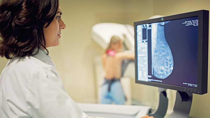
A mammogram is an X-ray of the breast that can be used to detect abnormalities. These breast screenings are one of the most effective ways to detect early signs of breast cancer.
When breast cancer is detected early from routine mammograms, it can provide the best chance for effective treatment.
While previous studies have shown that AI can improve radiologists’ detection of cancer in mammograms, the impact of AI on the search patterns of radiologists has been unknown until now.
Researchers used eye tracking to compare radiologists’ visual search patterns with and without AI support.
“By analysing this data, we can determine which parts of the mammograms the radiologist focuses on, and for how long, providing valuable insights into their reading patterns,” says joint first author Jessie Gommers, from the Department of Medical Imaging, Radboud University Medical Center in Nijmegen, Netherland.
The eye tracking system involved a small camera device with 2 infrared lights and a central camera. The device was then placed in front of the mammogram screen.
The camera picked up reflections of the radiologist’s eye from the infrared lights, allowing the research team to work out the exact coordinates the radiologist was looking at on the mammogram screen.
The eye tracking data showed radiologists with AI support spent more time examining the regions that contained lesions. Breast cancer detection accuracy was also higher with AI support.
“The results are encouraging,” Gommers says. “With the availability of the AI information, the radiologists performed significantly better.”
While radiologists with AI support spent more time examining regions with lesions, there was no evidence that these radiologists spent overall more time reading these mammograms, meaning cancer detection increased without extending reading time.
“Radiologists seemed to adjust their reading behaviour based on the AI’s level of suspicion: when the AI gave a low score, it likely reassured radiologists, helping them move more quickly through clearly normal cases,” Gommers says.
“Conversely, high AI scores prompted radiologists to take a second, more careful look, particularly in more challenging or subtle cases.”
The study involved 12 radiologists who read mammography examinations for 150 women, 75 with breast cancer and 75 without.
“Educating radiologists on how to critically interpret the AI information is key,” explains Gommers, who noted that an overdependence on AI could lead to unnecessary additional imagery or missed cancers.
While multiple studies have found the risk of incorrect AI information is relatively low when it comes to mammography interpretation, Gommers says it is important radiologists feel accountable for their own decisions.
Gommers and the research team are currently conducting additional studies, hoping to develop methods to predict if AI is uncertain about the decision.
“Overall, AI not only helped radiologists focus on the right cases but also directed their attention to the most relevant regions within those cases, suggesting a meaningful role for AI in improving both performance and efficiency in breast cancer screening.”